A Quantum Leap in Sensitivity
Traditional medical imaging modalities are fundamentally constrained by the classical physical limits of signal detection. Quantum sensing exploits the principles of superposition and entanglement to measure magnetic fields, electric fields, or temperature with unprecedented precision.
These sensors operate by meticulously controlling the quantum state of a system, such as an atomic vapor or a nitrogen-vacancy center in diamond, and observing how it is perturbed by the target physiological parameter.
This approach enables the detection of signals that were previously buried in noise, offering a path to imaging with higher spatial resolution, greater specificity for particular biomarkers, and at drastically reduced power levels compared to conventional techniques like MRI. The core advantage lies in moving beyond the standard quantum limit, a boundary that classical apparatus cannot surpass, thereby opening a new domain for diagnostic precision where minute biological processes become observable. This foundational shift is not merely an incremental improvement but represents a paradigm change in metrology for healthcare, allowing researchers to probe cellular function and neural activity with tools of exceptional accuracy and scale.
Beyond MRI The Promise of Diamond NV Centers
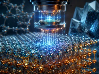
Among the most promising platforms for biomedical quantum sensing is the nitrogen-vacancy center in diamond. This atomic-scale defect acts as an optically addressable qubit with a spin state exquisitely sensitive to local magnetic fields.
Its quantum coherence can be maintained at room temperature, which is a critical advantage for practical clinical deployment over cryogenic alternatives.
Researchers are now developing NV-based microscopes and wearable sensors that can map neural action potentials, track individual magnetically-labeled cells, or image the microscopic flow of blood. The technique, known as diamond NV magnetometry, converts magnetic signals into detectable fluorescent changes, providing a non-invasive window into electrophysiology. Unlike conventional MRI, which measures net magnetization from vast ensembles of protons, NV centers can potentially detect the field from a single electron spin, pushing sensitivity to the theoretical limit. This capability allows for functional imaging of cellular networks and could revolutionize our understanding of metabolic processes at the sub-cellular level, offering a truly label-free method for observing ion channel dynamics and neurotransmitter release in real time.
| Platform | Key Measurand | Potential Medical Application | Current Resolution Frontier |
|---|---|---|---|
| Diamond NV Center | Magnetic Field (nanotesla) | Neuron Action Potential Mapping | Sub-micron / Single Cell |
| Atomic Vapor Cell | Magnetic Field (picotesla) | Fetal Magnetocardiography (fMCG) | Millimeter-scale |
| Superconducting Qubits | Microwave Photons | Molecular Spectroscopy | Single Photon Detection |
The transition from laboratory magnetometers to clinical devices requires significant engineering. Key challenges include integrating the diamond sensor with biological samples, minimizing environmental magnetic noise, and developing scalable fabrication for high-density sensor arrays. Progress in nanodiamond synthesis and waveguide integration is addressing these hurdles, paving the way for endoscopic and wearable quantum sensors.
Illuminating Brain Function with Entangled Light
Quantum optical imaging leverages non-classical light states, particularly entangled photon pairs, to see deeper into biological tissue with less damage. These techniques overcome fundamental limitations in classical optics, such as the diffraction limit and signal-to-noise barriers in scattering media.
By using photons that are intrinsically linked, measurements can be made with a precision that defies standard optical constraints. This enables high-resolution microscopy at greater depths, which is critical for observing the intricate circuitry of the living brain.
Applications like quantum optical coherence tomography and entangled two-photon microscopy provide clearer images of neural vasculature and cellular activity. They achieve this by distinguishing between signal photons and nnoise through quantum correlations, a method impervious to the random scattering that plagues traditional light-based imaging. The potential to monitor biophoton emission from metabolic processes or to track labeled molecules with unparalleled specificity is now within reach. This approach could transform functional brain imaging by providing direct, high-fidelity maps of synaptic communication and hemodynamic responses without the invasiveness of electrodes or the bulk of traditional scanners, marking a significant leap toward non-invasive neural circuit interrogation.
The inherent properties of entangled photons allow for lower illumination power, drastically reducing phototoxicity and enabling long-term observation of delicate biological processes. This makes it ideal for studying developmental biology or neurodegenerative diseases in model systems over extended periods. Researchers are actively working to translate these proof-of-concept lab systems into robust platforms suitable for preclinical research, which would provide neuroscientists with a transformative toolset.
What Are the Remaining Barriers to Clinical Adoption?
Despite the transformative potential, the path from laboratory demonstrators to routine clinical use is fraught with significant technical and practical hurdles. Quantum sensing systems often require exquisite isolation from environmental noise, precise temperature control, and complex optical alignment.
These conditions are difficult to maintain in a busy hospital setting outside a dedicated physics laboratory. The form factor of current devices is another major obstacle, as many setups are built on optical tables that are not portable or patient-accessible.
Developing compact, turnkey, and automated systems that can be operated by clinical staff without specialized physics training is a paramount engineering challenge. Material science must advance to produce cheaper, larger, and more consistent quantum-grade materials, such as diamonds with uniform NV center densities or efficient waveguide chips. Furthermore, the integration with existing clinical workflows and regulatory pathways for novel diagnostic devices presents a non-trivial challenge. Regulatory bodies like the FDA have limited experience evaluating the safety and efficacy of medical devices whose core function relies on quantum coherence, necessitating the development of new validation frameworks and performance standards. The high cost of prototype systems, driven by specialized lasers, cryogenics, and detection hardware, currently puts them beyond the reach of most healthcare institutions, demanding innovation to drive down costs through mass production techniques and simplified designs.
Beyond hardware, there is a critical need for robust data interpretation algorithms. The signals from quantum sensors are rich but complex, and extracting clinically relevant biomarkers requires advanced machine learning models trained on large, annotated datasets. The scarcity of such clinical quantum data is a catch-22 situation slowing progress. Collaborative efforts between quantum physicists, clinicians, and regulatory scientists are essential to build the necessary evidence base. Another subtle barrier is the therapeutic gap; a more sensitive diagnostic tool is only valuable if it leads to actionable clinical decisions or improved patient outcomes that are not possible with current technology. Demonstrating this clear clinical utility and cost-effectiveness will be the ultimate determinant of adoption, requiring longitudinal clinical studies that are time-consuming and expensive to conduct.
Significant investment is being directed toward overcoming these barriers, with a focus on creating hybrid systems that integrate quantum sensors with conventional imaging modalities. This strategy aims to provide an immediate, comparative benefit while the technology matures. Success hinges on achieving a balance between performance and practicality, ensuring that the quantum advantage is not nullified by operational complexity. The timeline for widespread clinical impact will depend on solving these multifaceted challenges, but the pace of innovation suggests that niche applications in neuroscience and cardiology could emerge within the next decade, paving the way for a broader revolution in precision medicine.
Charting the Future Diagnostic Landscape
The ultimate trajectory of quantum sensing in medicine points toward a new era of multimodal and multi-scale diagnostics. The integration of quantum sensors with established modalities like MRI, PET, and ultrasound will create hybrid platforms that leverage the unique strengths of each technology.
This convergence will enable simultaneous anatomical, functional, and molecular imaging, providing a comprehensive view of disease pathology that is currently unattainable.
For instance, a quantum-enhanced MRI could provide ultra-high-resolution structural images while simultaneously mapping real-time neural electrophysiology via diamond NV magnetometry, all within a single scanning session. The fusion of these data streams through advanced computational analytics will facilitate the discovery of novel biophysical biomarkers for early disease detection. In oncology, this might mean detecting the subtle magnetic signatures of tumor metabolism long before morphological changes are visible. For cardiology, it could enable the precise mapping of arrhythmogenic heart tissue with cellular precision. The shift from imaging anatomy to directly visualizing physiologcal function at the molecular level represents the core promise of this technological revolution, moving diagnostics from a descriptive to a predictive and mechanistic discipline. This evolution will necessitate parallel advances in computational power and AI-driven analysis to interpret the vast, complex datasets generated by these hyper-sensitive devices, transforming raw quantum signals into clinically actionable intelligence.
The development roadmap will likely see progressive miniaturization and specialization of sensors. Initial clinical applications will be in controlled environments for neuroscientific research and specialized cardiology. Wearable quantum sensors for continuous, ambulatory monitoring of biomarkers represent a longer-term but transformative goal. The following table outlines potential evolutionary stages for key quantum sensing platforms as they move toward clinical integration, highlighting the incremental steps from current proof-of-concept to future routine use.
| Technology Platform | Near-Term (5-7 years) | Mid-Term (7-12 years) | Long-Term Vision |
|---|---|---|---|
| Atomic Magnetometers | Specialized fetal MCG; brain function research. | Compact, helmet-style MEG for epilepsy monitoring. | Wearable arrays for continuous neurological & cardiac monitoring. |
| Diamond NV Centers | Ex vivo cellular imaging; biopsy analysis. | Endoscopic probes for surgical guidance; intraoperative microscopy. | In vivo subcellular metabolic imaging; distributed sensor networks. |
| Quantum Optical Imaging | Preclinical research in animal models. | Intraoperative margin assessment in oncology. | Non-invasive deep-tissue functional and metabolic microscopy. |
Beyond hardware, the ecosystem required for clinical quantum sensing is also taking shape. This includes the establishment of standardized protocols for sensor calibration, the creation of shared databases of quantum-biological signatures, and the training of a new generation of biomedical engineers and clinicians fluent in both quantum physics and medicine. Interdisciplinary collaboration will be the engine of progress, breaking down traditional silos between physics departments and medical schools.
Ethical and regulatory considerations will evolve alongside the technology. The unprecedented sensitivity of these devices raises important questions about data privacy, the potential for incidental findings, and the equitable access to such advanced, potentially costly ddiagnostics. Proactive engagement with these issues is essential to ensure the responsible development and deployment of quantum medical technologies. The economic model for these devices will also be critical, requiring demonstration of not just superior performance but also cost-effectiveness in improving patient outcomes and streamlining care pathways.
The journey from fundamental quantum research to hospital bedside is undoubtedly long and complex, yet the pace of advancement is accelerating. As material science, photonics, and nanofabrication continue to progress, the barriers of size, cost, and stability will gradually diminish. The first quantum sensors to achieve regulatory approval will likely address unmet clinical needs in neurology and cardiology, where their unique capabilities offer a clear diagnostic advantage. Their successful integration will then pave the way for broader adoption across other medical specialties, fundamentally altering the diagnostic paradigm and ushering in an age of truly personalized and predictive healthcare based on the most subtle signals of human physiology.