Mechanical Tension and Muscle Damage
The primary driver of exercise-induced hypertrophy is the development of high levels of mechanical tension within the muscle fibers. This tension, generated during resistance training, is detected by mechanosensors within the cell membrane and cytoskeleton. These sensors then translate the physical stimulus into a cascade of biochemical signals that promote anabolic processes.
Associated with high tension is the phenomenon of muscle damage, characterized by microscopic tears in the contractile elements and the sarcolemma. This damage, often leading to delayed onset muscle soreness, is not the primary driver of growth but serves as a potent signal. The subsequent inflammatory response and repair process recruit immune cells that clear debris and release signaling molecules that further activate satellite cells for proliferation.
The structural proteins desmin and titin, which maintain the integrity of the sarcomere, are particularly susceptible to disruption under heavy load. This disruption is a key event in initiating the remodeling process. Ultimately, the degree of tension and subsequent architectural disruption directly correlates with the magnitude of the anabolic response, provided adequate recovery is permitted.
Mechanotransduction pathways, including those mediated by focal adhesion kinase (FAK) and integrins, translate the mechanical stress of resistance exercise into biochemical signals that activate protein synthesis, with sensitivity to load and time under tension underscoring their central role. During recovery, protein degradation systems clear the wreckage, a seemingly catabolic but essential step that enables stronger rebuilding through hormesis. Crucially, specialized cells located between the basal lamina and muscle fiber membrane activate, proliferate, and fuse with existing fibers, donating new nuclei that expand the fiber’s long-term capacity for protein production and growth beyond its original genetic limits.
The following list outlines the sequential steps involved in the damage and repair cycle:
- Tension-Induced Microtears: High force disrupts sarcomeres and the extracellular matrix.
- Inflammatory Signaling: Immune cells like neutrophils and macrophages infiltrate the damaged area.
- Satellite Cell Activation: Local factors and systemic hormones trigger these cells to enter the cell cycle.
It is crucial to understand that the damage itself is not the goal, but a byproduct of the high forces required to stimulate adaptation. The focus should remain on progressively overloading the muscle with tension, as this is the most direct and effective method to ensure continuous growth. The inflammatory and repair processes will naturally follow when this primary stimulus is applied correctly.
Metabolic Stress and the Pump
A second major stimulus for muscle growth, often experienced during higher repetition training, is metabolic stress. This refers to the accumulation of metabolites such as lactate, inorganic phosphate, and hydrogen ions in the working muscle. The sensation of a intense "pump" or muscle swelling is the visible manifestation of this cellular environment.
The buildup of these byproducts is largely a result of performing exercises under conditions of restricted blood flow. When muscles contract intensely, intramuscular pressure exceeds capillary pressure, occluding venous return while arterial inflow may continue. This creates a hypoxic environment and traps metabolites within the muscle cells and interstitial space.
Metabolic stress is believed to contribute to hypertrophy through several mechanisms, including the promotion of anabolic hormone release. The local accumulation of metabolites can stimulate sensory nerves that, in turn, signal the pituitary gland to increase the output of growth hormone. This systemic hormonal spike is a classic response to high-repetition, short-rest training protocols.
Another significant effect is its role in promoting cellular swelling. The influx of water into the muscle cell, driven by the osmotic gradient created by metabolite accumulation, puts the cell membrane under tension. This mechanical stretch itself can be sensed as an anabolic signal, activating pathways involved in protein synthesis and inhibiting proteolysis.
The key physiological events induced by metabolite accumulation are listed below:
- Increased Hormone Release: Growth hormone and IGF-1 levels can be elevated systemically.
- Cellular Swelling: Cell hydration acts as a direct anabolic, anti-catabolic signal.
- Reactive Oxygen Species (ROS): Moderate ROS production from metabolic work can signal for adaptation.
Research indicates that the reactive oxygen species generated during metabolically demanding exercise also play a signaling role. While excessive ROS can be damaging, a controlled burst acts as a signal to upregulate antioxidant defenses and can sensitize the muscle to other growth signals. This adds another layer of complexity to how occlusion and metabolite buildup influence muscle remodeling.
The pump is more than a temporary cosmetic effect; it represents a distinct physiological state that complements the signals generated by high-threshold mechanical tension. While heavy lifting builds the foundational strength and disrupts contractile proteins, metabolic stress creates a systemic and local environment rich in anabolic hormones and cellular swelling, effectively supporting the repair and growth process initiated by the primary mechanical stimulus.
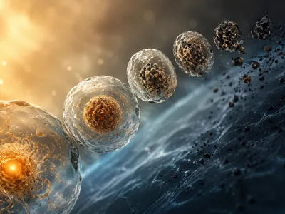
The Role of Satellite Cells in Repair
Satellite cells are quiescent myogenic stem cells located between the basal lamina and the sarcolemma of muscle fibers, responsible for repair and long-term skeletal muscle growth after damage or mechanical stress. They remain inactive until triggered by signals from injured tissue or high tension, including hepatocyte growth factor (HGF) released from the extracellular matrix and nitric oxide generated by shear stress. This activation is precisely controlled by signaling pathways and transcription factors, after which the cells proliferate to expand the pool of myogenic precursors needed for effective repair.
A significant portion of these proliferated cells will then differentiate and fuse with the existing damaged fiber. This fusion event is critical because it donates new myonuclei to the fiber, a concept often referred to as myonuclear domain theory. This addition expands the fiber's transcriptional capacity.
| Stage | Key Events | Regulatory Factors |
|---|---|---|
| Activation | Exit from quiescence, entry into cell cycle | HGF, FGF, Nitric Oxide, Notch signaling |
| Proliferation | Rapid division of myoblasts | MyoD, Myf5, mitogenic growth factors |
| Differentiation & Fusion | Alignment and fusion with existing fiber | Myogenin, MRF4, IGF-1, M-cadherin |
The newly donated myonuclei become permanent residents of the fiber, enabling it to sustain elevated rates of protein synthesis without overworking its original nuclei. This cellular addition is a fundamental reason why muscle fibers can grow substantially larger over time. Without this process, the fiber's genetic machinery would be a limiting factor for continued hypertrophy.
The efficiency of satellite cell activity appears to decline with age, a phenomenon linked to sarcopenia. However, resistance training has been shown to rejuvenate some of this capacity, even in older populations. This underscores the importance of consistent mechanical loading throughout the lifespan to maintain both muscle mass and the regenerative potential of the stem cell pool.
The interplay between satellite cells and the local microenvironment, or niche, is crucial. Inflammatory cells, such as M2 macrophages, play a supportive role by clearing debris and secreting cytokines that promote satellite cell proliferation and differentiation. This coordinated cellular conversation ensures that the repair process is both efficient and leads to a net gain in structural protein.
How Hormones Influence Protein Synthesis
The endocrine system strongly modulates muscular adaptation to resistance training, as hormones like testosterone, growth hormone (GH), and insulin-like growth factor-1 (IGF-1) drive anabolic activity by regulating muscle protein synthesis. Testosterone binds to androgen receptors in muscle cells, moves to the nucleus, and alters gene transcription to increase contractile protein production while reducing breakdown. Meanwhile, the GH/IGF-1 axis exerts much of its effect through locally produced IGF-1—particularly the mechano-growth factor (MGF) variant— which responds to mechanical load and plays a central role in satellite cell activation.
The actual signaling cascade initiated by IGF-1 binding to its receptor is a classic example of a growth pathway. It activates the PI3K-Akt-mTOR pathway, which is often described as the central hub for regulating protein synthesis. Akt directly phosphorylates and activates mTOR, which then orchestrates the assembly of the translational machinery.
Cortisol, a glucocorticoid hormone, represents the primary catabolic counterpart to these anabolic signals. While essential for metabolism and the stress response, chronically elevated cortisol can suppress protein synthesis and accelerate protein degradation, creating an environment unfavorable for hypertrophy. The balance between anabolic and catabolic hormones, rather than the absolute level of any single hormone, often dictates the net gain in muscle tissue.
Acute resistance exercise temporarily elevates the levels of anabolic hormones like testosterone and GH. This post-exercise hormonal spike is thought to contribute to the overall adaptive environment, although its necessity for hypertrophy in well-trained individuals is a topic of ongoing investigation. The local production of autocrine and paracrine factors within the muscle may play an equally, if not more, significant role.
Nutrition, Recovery, and Optimal Timing
The anabolic stimuli from tension and hormonal signaling are rendered ineffective without the fundamental building blocks and sufficient recovery periods. Nutrient availability and sleep quality directly dictate whether the cellular environment favors synthesis or degradation.
Dietary protein provides the essential amino acids, particularly leucine, that serve as both substrates and signaling molecules for muscle protein synthesis. Consuming a sufficient dose of high-quality protein post-exercise effectively "turns on" the translational machinery. Spreading protein intake evenly across meals appears superior to skewed distribution for maintaining a positive nitrogen balance.
Achieving a net positive energy surplus is often necessary for significant hypertrophy, as the process of building new tissue is energetically costly. While a caloric deficit can permit some growth in untrained individuals, advanced trainees typically require additional energy to fuel the heightened rates of synthesis. Carbohydrates play a crucial role here, not only by replenishing glycogen but also by spiking insulin, which has a permissive effect on protein synthesis and helps to mitigate protein breakdown. Fats are equally vital for maintaining hormonal health, including the production of endogenous testosterone.
The relationship between specific nutrients and their direct physiological impact on muscle remodeling is outlined below. This table summarizes key dietary components and their primary functions in the hypertrophic process.
| Nutrient/Component | Primary Role in Hypertrophy |
|---|---|
| Complete Proteins (e.g., whey, egg) | Provide essential amino acids, especially leucine, to directly stimulate MPS. |
| Carbohydrates | Replenish glycogen stores and spike insulin, creating a more anabolic hormonal milieu. |
| Dietary Fats | Support basal hormone production, including testosterone, and overall cellular health. |
| Creatine Monohydrate | Enhances phosphocreatine stores, allowing for higher training volume and intensity. |
Sleep represents a critical, often underestimated, period of recovery where the majority of growth hormone release and protein synthesis occurs. Poor sleep quality elevates cortisol and blunts the anabolic response to feeding and training. The primacy of sleep for hormonal optimization cannot be overstated, as it sets the stage for all other recovery processes.
Beyond sleep and nutrition, specific behavioral and training practices can significantly influence recovery kinetics. Implementing these strategies helps ensure consistent progress and reduces the risk of overtraining.
- Active Recovery: Low-intensity movement on rest days enhances blood flow and nutrient delivery to muscle tissue.
- Stress Management: Chronic psychological stress elevates cortisol, directly antagonizing anabolic signaling pathways.
- Periodized Deloads: Scheduled reductions in training volume or intensity allow for complete systemic recovery and joint repair.
The concept of nutrient timing has evolved from a rigid post-workout "anabolic window" to a more nuanced understanding of total daily intake. While consuming protein shortly after training can be beneficial, particularly for older adults, the total amount and distribution across the day remain the dominant factors. Ensuring that high-quality protein is consumed at regular intervals, especially before sleep to support overnight MPS, is a practical and evidence-based strategy for maximizing growth.