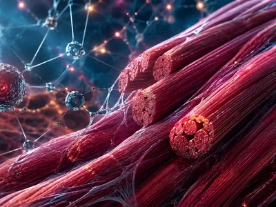
The Genome in Disarray
Cellular aging is driven by the gradual accumulation of DNA damage, including single-strand breaks, base modifications, and double-strand fractures, which eventually overwhelm repair mechanisms. This genomic instability activates the DNA damage response (DDR), halting the cell cycle and promoting inflammatory secretions that affect neighboring cells.
Alterations in the epigenetic landscape exacerbate aging, as aberrant DNA methylation and histone modifications lead to transcriptional noise and loss of cellular identity. Additionally, loosening of heterochromatin allows transposable elements to reactivate, inserting into critical genomic regions and further destabilizing the genome.
Persistent DDR signaling induces cellular senescence, creating a pro-inflammatory microenvironment through the senescence-associated secretory phenotype (SASP). These molecular derangements form a feedback loop: inflammation increases DNA damage, while epigenetic drift reduces repair enzyme expression, accelerating functional decline and cellular aging.
- Oxidative base modifications 8-oxoG
- Telomere-associated foci (TAFs) DNA-SCARS
- Lamin B1 depletion Nuclear Envelope
- Mitochondrial DNA deletions mtDNA
Telomeres: The Fraying Ends
Linear chromosomes face an inherent end-replication problem, where each cell division results in the loss of terminal DNA sequences. These protective caps, known as telomeres, consist of repetitive TTAGGG repeats bound by the shelterin protein complex.
When telomeres shorten to a critical length, they lose shelterin protection and are recognized as double-strand breaks. This forces the cell into either replicative senescence or apoptosis, effectively limiting the proliferative capacity of somatic tissues.
The enzyme telomerase counteracts this attrition by adding telomeric repeats, yet it is silenced in most human somatic cells. Only germ cells, stem cells, and certain immune cells maintain sufficient telomerase activity to sustain long-term proliferation, creating a stark divide in replicative potential across cell types.
Emerging evidence reveals that telomere dysfunction extends far beyond simple shortening. Telomere uncapping can occur even at moderate lengths due to oxidative damage to the telomeric DNA or depletion of shelterin components such as TRF2. This uncapping triggers a robust DDR that is indistinguishable from that caused by overt genomic breaks. Moreover, dysfunctional telomeres contribute to systemic aging through cell-non-autonomous mechanisms; senescent cells with short telomeres secrete SASP factors that propagate inflammation to distal tissues, thereby accelerating aging throughout the organism.
| Telomere Maintenance Strategy | Cell Type / Context | Consequence for Aging |
|---|---|---|
| High telomerase activity | Germline, stem cells, activated lymphocytes | Prolonged replicative capacity |
| Alternative lengthening of telomeres (ALT) | Certain cancer cells, some mesenchymal stem cells | Can bypass senescence but risk malignant transformation |
| Progressive attrition | Most somatic cells | Replicative senescence and tissue dysfunction |
Interventions that modulate telomere dynamics, such as telomerase activation in regenerative contexts or clearance of telomere-damaged senescent cells, are now being explored as therapeutic strategies to delay age-related pathologies. The delicate balance between preserving replicative potential and suppressing cancer remains a central challenge in this field.
When Cells Stop Dividing
Once celebrated solely as a tumor-suppressive mechanism, cellular senescence is now understood as a major driver of tissue aging. This state of permanent cell cycle arrest is triggered by various stressors, including telomere erosion, DNA damage, and oncogene activation.
Senescent cells evade apoptosis and accumulate in aged organisms, creating a toxic microenvironment through the senescence-associated secretory phenotype (SASP). The SASP includes interleukins, chemokines, growth factors, and matrix-degrading enzymes that disrupt tissue architecture and spread inflammation to neighboring cells.
A critical aspect of senescence is its heterogeneous nature. Depending on the inducing stimulus and cell type, senescent cells can exhibit markedly different secretory profiles and metabolic rewiring. Some senescent cells adopt a pro-fibrotic secretome, while others primarily drive immune cell recruitment, illustrating that senescence is not a uniform endpoint but a dynamic, context-dependent state.
The chronic presence of these cells has been causally linked to osteoarthritis, atherosclerosis, neurodegeneration, and metabolic dysfunction. Genetic or pharmacological clearance of senescent cells—an approach known as senolysis—has been shown to extend healthspan and mitigate age-related decline in preclinical models, validating senescent cells as therapeutic targets.
-
Biomarkerp16INK4aCyclin-dependent kinase inhibitor; a gold-standard senescence marker
-
TherapySenolyticsDrugs that selectively eliminate senescent cells (e.g., dasatinib + quercetin)
-
EffectorsSASP factorsIL-6, IL-1β, CXCL1, MMPs; paracrine drivers of inflammaging
Power Failure and Garbage Accumulation
Mitochondrial dysfunction lies at the intersection of metabolic decline and cellular aging. As mitochondria accumulate oxidative damage to their DNA and membranes, they shift from efficient ATP production toward increased reactive oxygen species (ROS) emission, creating a self-perpetuating cycle of injury.
This bioenergetic deterioration is accompanied by a collapse of proteostasis—the cellular quality-control network that maintains protein folding and degradation. Impaired autophagy and ubiquitin-proteasome system activity allow misfolded and aggregated proteins to accumulate, forming inclusion bodies that interfere with normal cellular functions.
The interplay between mitochondrial stress and defective proteostasis is particularly damaging in post-mitotic cells such as neurons and cardiomyocytes. Here, lipofuscin—an indigestible aggregate of oxidized proteins and lipids—accumulates over time, physically crowding the cytosol and further impeding lysosomal function. This vicious cycle of metabolic failure and waste accumulation is a unifying feature of many age-related degenerative disorders.
| Pathological Feature | Primary Cause | Consequence |
|---|---|---|
| Mitochondrial DNA deletions | Replication errors & ROS damage | Impaired electron transport chain, reduced ATP |
| Lipofuscin accumulation | Incomplete lysosomal degradation | Cytoplasmic crowding, autophagy blockade |
| Aggregated proteins | Proteasome/autophagy decline | Proteotoxic stress, cellular dysfunction and apoptosis |
| Mitochondrial permeability transition | Calcium overload & oxidative stress | Release of pro-apoptotic factors, cell death |
Emerging strategies to restore cellular energy balance and clearance capacity include pharmacological activation of mitophagy, boosting NAD⁺ levels to enhance sirtuin-dependent mitochondrial quality control, and employing proteostasis regulators that refold or degrade toxic aggregates. Such interventions aim to disrupt the nexus of power failure and garbage accumulation, thereby preserving tissue function across the lifespan.