The Sleep-Wake Symphony and Synaptic Homeostasis
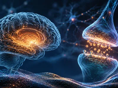
The brain’s neural architecture is not static but undergoes dynamic change across the states of wakefulness and sleep. A core hypothesis posits that synaptic homeostasis is a fundamental function of sleep, counterbalancing the net increase in synaptic strength that occurs during daily learning. This process is likened to a symphony where neural activity during slow-wave sleep orchestrates a widespread downscaling of synapses, preserving overall energetic efficiency and network stability. By reducing the metabolic cost of maintaining potentiated connections, sleep prevents neural saturation and ensures cognitive resources remain available for new experiences.
The molecular and electrophysiological signatures of this downscaling are now increasingly understood. During deep, non-REM sleep, the synchronized slow oscillations and associated delta waves create a permissive state for synaptic renormalization. These large-scale brain rhythms facilitate a global reduction in synaptic weight that is proportional to the strength gained during prior wakefulness. Crucially, this does not erase memory traces but rather sharpens the signal-to-noise ratio, enhancing the most salient connections while weakening less significant ones. The intricate dance between long-term potentiation (LTP) during wake and long-term depression (LTD) during sleep forms the basis of this homeostatic regulation. Disruptions in this cycle, therefore, do not merely cause fatigue but fundamentally impair the brain’s capacity to maintain an adaptable and efficient neural network, highlighting sleep’s role as a non-negotiable biological requirement for cognitive integrity.
Synaptic Pruning
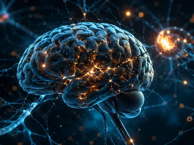
Beyond general downscaling, sleep facilitates a more selective editing of neural circuits known as synaptic pruning. This targeted elimination of weaker connections is essential for refining brain networks, particularly during developmental critical periods but also throughout adulthood. Pruning works in concert with activity-dependent strengthening to sculpt the connectome, removing redundant or inefficient pathways.
The mechanisms of sleep-dependent pruning involve specific molecular tags. Proteins such as complement factors C1q and C3 are upregulated during wakefulness and mark underutilized synapses for removal. Sleep, particularly the deep stages, then activates microglial cells, the brain’s resident immune cells, to engulf and clear these tagged connections. This process is not random but is guided by neural activity patterns from the preceding day.
Sleep therefore transforms a potentially harmful inflammatory pathway into a precision tool for neural optimization. The following list outlines key functional outcomes of effective synaptic pruning during sleep:
- Enhanced computational efficiency by streamlining neural pathways.
- Improved signal fidelity and separation of memory engrams.
- Prevention of hyperexcitability and excessive cortical connectivity, which are risk factors for disorders.
- Provision of physical space and metabolic resources for subsequent new synaptic formation.
This nightly editing is critical for cognitive functions like executive control and flexible thinking. Without it, the brain’s wiring would become cluttered and noisy, hindering optimal performance. The precise coordination between neuronal activity, molecular signaling, and glial function underscores the biological complexity of what was once considered a passive state, revealing sleep as an active period of essential network maintenance and refinement.
Memory Consolidation Pathways
Sleep orchestrates the stabilization and integration of memories through distinct neurophysiological events. This process, known as systems consolidation, involves a dynamic dialog between the hippocampus and neocortex. During slow-wave sleep, the reactivation of hippocampal memory traces, called sharp-wave ripples, drives the transfer of information to long-term storage sites.
The coordination of different sleep stages creates an optimal environment for various memory types. Non-REM sleep, with its slow oscillations and spindles, is particularly crucial for declarative memories—facts and events. These brain rhythms facilitate the safe replay and redistribution of hippocampal-dependent memories to the cortical networks where they become more stable and integrated with existing knowledge.
In contrast, REM sleep, characterized by rapid eye movements and high cortical activity, appears vital for procedural and emotional memory processing. The cholinergic environment and theta rhythms during REM may promote synaptic plasticity in circuits involved in skills and emotional regulation. The following table summarizes the primary sleep stages associated with different memory systems:
| Sleep Stage | Dominant Oscillations | Associated Memory Type | Proposed Function |
|---|---|---|---|
| Slow-Wave Sleep (N3) | Slow Oscillations, Delta Waves, Sleep Spindles | Declarative (Semantic & Episodic) | Systems Consolidation, Hippocampal-Neocortical Dialogue |
| REM Sleep | Theta Rhythms, Ponto-Geniculo-Occipital Waves | Procedural, Emotional, Spatial | Synaptic Plasticity, Emotional Integration |
| Stage N2 Sleep | Sleep Spindles, K-Complexes | Motor Learning, Factual Recall | Local Synaptic Stabilization |
This staged process is not merely about preservation but also involves active transformation and integration. Memories are often restructured, with irrelevant details fading and core concepts becoming more salient. This reorganization fosters insight and creative problem-solving upon waking. The role of sleep spindles is especially noteworthy, as their density correlates with intelligence measures and learning success, acting as a key mechanism for thalamocortical circuit plasticity.
Memory consolidation relies on specific neuromodulatory shifts. The low acetylcholine levels during non-REM sleep permit hippocampal replay, while the high acetylcholine during REM may facilitate cortical plasticity. The key molecular events supporting this include:
- Increased expression of plasticity-related genes such as BDNF (Brain-Derived Neurotrophic Factor).
- Protein synthesis-dependent long-term potentiation occurring in cortical networks.
- Calcium-dependent molecular cascades that strengthen reactivated synaptic connections.
How Does Sleep Enhance Learning?
Sleep enhances learning capacity through preparatory mechanisms that restore synaptic plasticity. A rested brain exhibits greater potential for long-term potentiation (LTP), the cellular basis of learning, compared to a sleep-deprived one. This restoration involves both the clearance of metabolic byproducts that inhibit plasticity and the replenishment of neurotransmitter vesicles.
Prior sleep optimizes the neural substrate for encoding new information. It resets the network to a baseline state where new synaptic potentiation can occur efficiently. Without this reset, neural circuits operate at a saturated level, diminishing their ability to register new experiences. This explains why learning a new skill is more effective after sleep than before it.
Sleep after learning, as discussed, consolidates memories. Sleep before learning, however, is equally critical for preparing the hippocampus to acquire novel information. This dual function underscores that sleep is not a mere post-learning luxury but an integral part of the entire learning cycle. Sleep deprivation severely impairs hippocampal function, reducing the ability to form new episodic memories by up to forty percent in some studies.
The benefits extend beyond simple recall to higher-order cognitive integration. Sleep fosters the extraction of gist, the discovery of hidden patterns, and the ability to make nnovel inferences from learned material. This cognitive enhancement suggests sleep promotes a reorganization of memory representations, allowing for more flexible and creative use of knowledge.
The Glymphatic System and Cellular Detox
A groundbreaking discovery in neuroscience is the glymphatic system, a brain-wide waste clearance network that becomes highly active during sleep. This system utilizes the perivascular spaces surrounding cerebral blood vessels to facilitate the exchange of cerebrospinal fluid (CSF) and interstitial fluid (ISF), effectively washing the brain parenchyma.
The driving force behind this cleansing process is the synchronized activity of neural and glial cells during deep non-REM sleep. Astrocytic endfeet regulate the flow, and the expansion of the interstitial space by up to sixty percent during slow-wave sleep allows for efficient fluid movement. This physiological state creates a powerful detoxification cycle that removes accumulated metabolic waste products from the day's neural activity.
One of the most critical functions of this nightly wash is the clearance of neurotoxic proteins, including amyloid-beta and tau, which are implicated in neurodegenerative diseases. The efficiency of amyloid-beta removal during sleep is significantly higher compared to wakefulness. The table below outlines key characteristics and functions of the glymphatic system:
| Component | Function | Sleep-Associated Change |
|---|---|---|
| Astrocytic Aquaporin-4 (AQP4) Channels | Regulates CSF-ISF exchange along perivascular pathways | Polarization and function are optimized during slow-wave sleep |
| Interstitial Space Volume | Conduit for waste-laden fluid movement | Dramatically increases, enhancing convective flow |
| Arterial Pulsatility | Drives fluid movement through the brain | Couples with slow oscillations to boost glymphatic influx |
| Cerebrospinal Fluid (CSF) Inflow | Delivers fresh fluid and removes solutes | Increases substantially, peaking during deep sleep phases |
This process is not a simple passive diffusion but an active, energy-dependent clearance mechanism fundamentally tied to the electrical rhythms of the sleeping brain. The synchronization of neuronal slow oscillations, blood flow, and breathing rhythms appears to pump CSF through the brain, linking neural activity directly to metabolic homeostasis. Consequently, disruptions in sleep architecture or quality can impair this essential maintenance, allowing potentially harmful proteins to aggregate.
The implications for long-term brain health are profound. Chronic sleep disruption is now recognized as a major modifiable risk factor for cognitive decline. By compromising the glymphatic system, poor sleep may accelerate the pathological accumulation of proteins associated with Alzheimer's disease and other dementias. The primary waste products cleared by the glymphatic system during sleep include:
- Amyloid-beta peptides Primary Alzheimer's-associated protein
- Tau protein aggregates Linked to neurofibrillary tangles
- Lactate and other metabolic byproducts Result of daily neuronal activity
- Inflammatory cytokines Potential neuroinflammatory mediators
Therefore, sleep serves as a critical period for cellular detoxification and metabolic reset, protecting the structural and functional integrity of neural networks. This recently elucidated system provides a compelling physiological explanation for the non-negotiable requirement of sleep and its deep connection to neurological resilience over the lifespan.
Sleep Deprivation’s Neural Consequences
Acute and chronic sleep loss precipitates a cascade of detrimental effects on neural connectivity and cognitive function. The disruption of synaptic homeostasis leads to a state of synaptic sturation, where neurons struggle to potentiate further in response to new stimuli. This manifests as impaired learning capacity and reduced hippocampal activity during memory encoding tasks.
Furthermore, the essential processes of synaptic pruning and memory consolidation are severely curtailed. Without the selective weakening of irrelevant connections, neural circuits become noisy and inefficient. The reactivation and redistribution of memory traces are disrupted, leaving recently acquired information vulnerable to interference and loss.
Sleep deprivation also cripples the glymphatic clearance mechanism, allowing neurotoxic metabolites to accumulate in the interstitial space. This creates a neuroinflammatory environment that can damage neurons and glial cells over time. The combined impact on synaptic plasticity, network refinement, and cellular health underscores why cognitive deficits from sleep loss are so pervasive.
Long-term consequences extend to the structural level, with studies showing alterations in white matter integrity and reduced cortical volume in chronically sleep-deprived individuals. The brain’s ability to maintain energy balance is also compromised, as the restorative metabolic processes that occur during sleep are missed. Ultimately, persistent sleep disruption represents a significant risk factor for a spectrum of neurological and psychiatric disorders, from major depression to neurodegenerative disease, highlighting the indispensable role of sleep in preserving the very architecture of the mind.