From Cartography to Connectomics
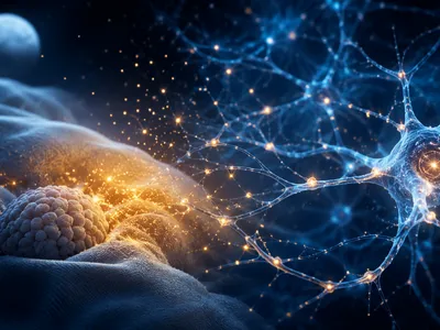
The fundamental quest to understand the brain's structure has evolved from macroscopic anatomical sketches to the molecular-scale reconstruction of its wiring. Classical brain cartography, pioneered by Brodmann and others, provided a foundational cytoarchitectonic map based on cellular organization. This histological approach, however, lacked functional and connectional specificity. The field's paradigm shifted with the emergence of connectomics, which aims to comprehensively catalog neural connections—the connectome—as a complete wiring diagram of the brain.
Modern connectomics leverages advanced imaging techniques like serial section electron microscopy and high-throughput light microscopy to visualize synaptic-level details across large tissue volumes. This data-intensive endeavor requires not only technological prowess but also sophisticated computational tools for image segmentation, neural tracing, and network analysis. The primary goal is to move beyond static maps and understand how specific neural circuits give rise to behavior, perception, and cognition, fundamentally bridging the gap between structure and function in neuroscience.
Lighting Up the Brain The Optogenetics Revolution
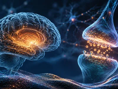
A pivotal breakthrough in functional brain mapping is optogenetics, which provides unprecedented cell-type-specific control over neuronal activity. By integrating microbial opsins, light-sensitive ion channels, into defined neuronal populations, researchers can precisely excite or inhibit neurons with millisecond precision using pulses of light. This technique transformed causal interrogation of neural circuits from a correlative science into an exact experimental paradigm.
The mapping power of optogenetics lies in its ability to establish direct links between targeted neural activity and behavioral outcomes. When combined with fiber photometry or miniature microscopes, it allows for simultaneous manipulation and observation of activity in freely behaving animals. This closed-loop experimentation has been instrumental in deconstructing circuits underlying reward, fear, social behavior, and motor control.
More recently, the development of multicolor optogenetic tools enables independent control of multiple neural populations within the same region, revealing how coordinated ensemble activity encodes information. The spatial precision of optogentics has been further enhanced by patterned illumination techniques, such as holographic photostimulation, which can activate neurons in user-defined, three-dimensional patterns to simulate naturalistic neural codes.
The convergence of optogenetics with other modalities, like fMRI (opto-fMRI) or electrophysiology, creates a powerful multimodal mapping platform. This allows scientists to observe both the local cellular effects and the global, brain-wide consequences of precisely perturbing a specific circuit node, offering a comprehensive view of network dynamics and causal hierarchies within the brain's interconnected architecture.
The Symphony of Neuronal Recordings
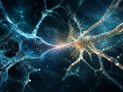
Complementing optogenetic manipulation, advanced electrophysiological recording technologies now capture the brain's intricate electrical symphony with astonishing resolution. High-density neural probes, such as Neuropixels, can simultaneously monitor hundreds to thousands of individual neurons across multiple brain regions in behaving animals.
This massive parallel recording generates unprecedented datasets, revealing complex patterns of coordinated ensemble activity that underlie cognition.
The analysis of these high-dimensional datasets has necessitated sophisticated computational approaches. Techniques like population vector analysis and dimensionality reduction (e.g., t-SNE, UMAP) allow researchers to visualize how neural representations evolve over time during tasks. Furthermore, the application of machine learning classifiers to neural activity can decode an animal's intentions, perceptions, or decisions with increasing accuracy, effectively "reading out" brain states. This convergence of large-scale recording and computational analysis is crucial for testing theoretical models of neural coding, moving beyond single-neuron responses to understand how information is distributed and processed across vast, interconnected networks. The ultimate goal is to decipher the neural syntax—the rules governing how the collective activity of neurons gives rise to coherent functions.
- High-Density Silicon Probes (e.g., Neuropixels 2.0): Enable recording from >10,000 sites, spanning deep brain structures with single-cell resolution.
- Calcium Imaging Miniaturization: Head-mounted microscopes (miniscopes) allow calcium imaging of thousands of neurons in freely moving subjects, linking molecular activity to behavior.
- Flexible Bioelectronic Interfaces: Polymer-based, tissue-conformable electrodes reduce immune response and enable stable chronic recordings for longitudinal studies of learning and plasticity.
Computational Giants Mapping the Invisible
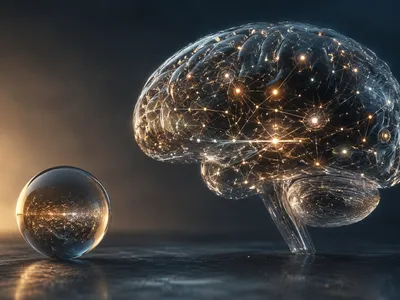
The deluge of data from connectomics, optogenetics, and massive recordings is futile without the computational frameworks to integrate and interpret it. Computational neuroscience and artificial intelligence have become indispensable partners in modern brain mapping, creating in silico models that predict function from structure. These models simulate network dynamics based on anatomical connectivity, testing hypotheses about signal propagation and information flow.
A prime example is the development of whole-brain simulation platforms, like the Blue Brain Project, which aim to reconstruct and simulate rodent brain regions at cellular detail. These efforts rely on powerful algorithms to infer missing biological parameters and to translate structural maps into functional models, revealing emergent properties not evident from anatomy alone.
Machine learning, particularly deep learning, is revolutionizing the analysis of brain mapping data. Convolutional neural networks automate the labor-intensive segmentation of electron microscopy images for connectomics, while graph neural networks analyze the resulting connectomes to identify crucial network hubs and motifs. Simultaneously, AI is used to model the brain's own computations, with artificial neural networks providing a new lens to understand how hierarchical sensory processing or reinforcment learning might be implemented biologically. This bidirectional flow—using brain data to inspire AI and using AI to analyze the brain—accelerates discovery and bridges the gap between observable neural activity and abstract cognitive theory.
| Computational Approach | Primary Application in Brain Mapping | Key Outcome |
|---|---|---|
| Network Theory & Graph Analysis | Analyzing structural and functional connectomes | Identifies hub regions, modularity, and resilience properties of brain networks. |
| Biophysical Neural Simulation | Predicting dynamics from cellular properties and connectivity | Reveals how microcircuit motifs generate oscillations and signal integration. |
| Deep Learning (Computer Vision) | Automated image segmentation for connectomics | Enables large-scale, high-accuracy reconstruction of neural wiring from EM data. |
| Representational Learning | Analyzing high-dimensional neuronal recording data | Decodes cognitive states and reveals latent variables driving neural activity. |
A Dynamic Atlas Implications for Mind and Machine
The convergence of these mapping technologies is yielding not a static diagram but a dynamic, multi-scale atlas of the living brain. This paradigm shift recognizes that the connectome is not fixed; it exhibits experience-dependent plasticity, modulated by neuromodulators and changing with behavioral states. Modern approaches now aim to capture this dynamism, mapping how functional connectivity reconfigured during tasks, sleep, or in neurological disorders, providing a more accurate representation of the brain's operational logic.
For neuroscience, this dynamic atlas offers a foundational framework for mechanistically understanding brain disorders. It enables the identification of specific circuit dysfunctions underlying conditions like depression, schizophrenia, or Parkinson's disease, moving beyond symptomatic diagnosis towards circuit-based nosology. This precision facilitates the development of targeted neuromodulation therapies, such as closed-loop deep brain stimulation, which can adapt to the brain's real-time activity to correct pathological oscillations.
The implications extend powerfully into artificial intelligence and brain-computer interfaces. The principles of efficient, robust, and adaptive network organization discovered in biological brains are inspiring the next generation of neuromorphic computing architectures. These chips, designed to mimic the brain's parallel processing and synaptic plasticity, promise breakthroughs in energy-efficient AI. Conversely, detailed brain maps are critical for designing high-fidelity BCIs. By understanding the coordinate system and population codes of motor or speech cortex, decoders can translate neural activity into device control with unprecedented nuance and reliability, potentially restoring lost functions. This reciprocal dialogue between neuroscience and engineering is accelerating progress in both fields, blurring the boundary between biological and artificial intelligence and pushing the frontiers of what is possible in understanding and interfacing with the mind.
The grand challenge of brain mapping transcends mere anatomical cataloging. It seeks to establish a causal, predictive model of brain function—a computational framework that can simulate how sensory input propagates through a personalized connectome, is processed by dynamic neural ensembles, and culminats in thought and action. Achieving this would represent a pinnacle of scientific understanding, with profound implications for medicine, technology, and our fundamental conception of self. The path forward requires continued integration across scales and disciplines, leveraging each breakthrough to refine this ever-evolving atlas of human consciousness.