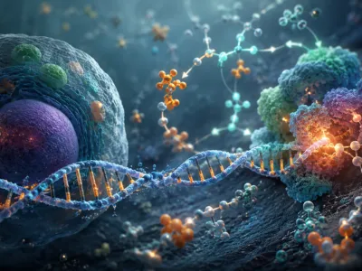
Metabolic Pathways in Flux
Dietary restriction triggers a major reorganization of hepatic gluconeogenesis and ketogenesis, shifting energy use from glucose to fatty acid oxidation. This adaptation is regulated by the AMP-activated protein kinase (AMPK) complex, which functions as a cellular energy sensor and suppresses anabolic pathways during caloric deficit.
The transition from fed to fasted states alters redox homeostasis within mitochondria, modulating key enzymes like carnitine palmitoyltransferase 1 (CPT1). At the same time, suppressed insulin signaling lowers malonyl-CoA levels, allowing unhindered fatty acid import for β-oxidation and ketone body production, providing alternative fuel for extrahepatic tissues.
Insulin's Gatekeeper Role
Insulin orchestrates whole-body energy partitioning by directly inhibiting adipose triglyceride lipase (ATGL) and hormone-sensitive lipase (HSL), thereby halting lipolysis during the postprandial period.
In the context of energy restriction, declining insulin concentrations relieve this inhibition, allowing unopposed mobilization of stored triglycerides into the circulation as free fatty acids.
The molecular cascade downstream of insulin receptor activation involves the phosphoinositide 3-kinase (PI3K)/Akt pathway, which phosphorylates and inactivates forkhead box O1 (FOXO1), a transcription factor essential for gluconeogenic gene expression. Prolonged low insulin states consequently promote autophagy-mediated lipid droplet turnover within adipocytes, fundamentally altering adipose tissue morphology and enhancing metabolic flexibility during sustained caloric deficit.
The following key hormonal regulators illustrate the integrated control over lipid mobilization:
- Insulin – Suppresses lipolysis via Akt-mediated phosphorylation of lipases
- Glucagon – Stimulates lipolysis through cAMP-dependent protein kinase A (PKA)
- Catecholamines – Activate β-adrenergic receptors to drive hormone-sensitive lipase phosphorylation
These counter-regulatory signals converge on perilipin-1, a lipid droplet scaffold protein whose phosphorylation state dictates accessibility of stored triglycerides to cytosolic lipases.
The Metabolic Switch: Ketosis Explained
Hepatic ketogenesis increases as glycogen stores are depleted and insulin levels drop, shifting the liver from glucose consumption to fatty acid oxidation. This transition produces a surge in β-hydroxybutyrate and acetoacetate, which act as efficient energy sources for neurons and cardiomyocytes.
The rate-limiting enzyme is 3-hydroxy-3-methylglutaryl-CoA synthase 2 (HMGCS2), whose expression is inhibited by insulin and stimulated by glucagon through PPARα. Accumulating acetyl-CoA from fatty acid β-oxidation is redirected from the TCA cycle toward ketone synthesis, a process enhanced by reduced malonyl-CoA levels that permit mitochondrial fatty acid uptake, sustaining a ketotic state vital for energy balance during prolonged dietary restriction.
Macronutrient Signaling and Satiety
Dietary protein and fiber exert pronounced effects on appetite regulation through distinct gut-brain signaling axes, modulating release of anorexigenic hormones such as glucagon-like peptide-1 (GLP-1) and peptide YY (PYY).
Fermentable carbohydrates generate short-chain fatty acids that activate free fatty acid receptors on enteroendocrine L-cells, amplifying satiety signaling while suppressing ghrelin secretion from gastric cells.
The convergence of these signals at the hypothalamic arcuate nucleus determines net food intake through nuanced integration of hormonal and nutrient-derived stimuli.
Beyond gut-derived peptides, amino acid sensing mechanisms in the hypothalamus directly influence neuropeptide Y/agouti-related peptide (NPY/AgRP) neurons, creating a feedback loop that adjusts hunger perception based on circulating essential amino acid profiles. This intricate molecular dialogue underscores why protein-rich dietary patterns consistently demonstrate superior satiety outcomes compared to isoenergetic meals dominated by simple carbohydrates, a phenomenon rooted in the differential activation of mammalian target of rapamycin (mTOR) signaling within metabolic sensing neurons, representing one of the many cutting-edge biochemistry applications in nutrition science.
Thermic Effect of Food: A Hidden Factor
The thermic effect of food (TEF) represents the energy expended during digestion, absorption, and disposal of ingested nutrients, varying substantially by macronutrient composition, showcasing the direct impact of biochemistry in everyday life.
Protein elicits the highest TEF, ranging from 20 to 30 percent of its caloric content, whereas carbohydrates and fats yield TEF values of 5 to 10 percent and 0 to 3 percent, respectively. This disparity arises from the adenosine triphosphate (ATP) demands of ureagenesis, gluconeogenesis, and peptide bond synthesis, which collectively impose a substantial metabolic overhead. Consequently, dietary patterns enriched with protein and complex carbohydrates can create a measurable advantage in daily energy expenditure, a factor often overlooked in conventional calorie-restricted regimens.
The following table illustrates macronutrient-specific contributions to diet-induced thermogenesis:
| Macronutrient | Thermic Effect Range (% of intake) | Primary Metabolic Pathway |
|---|---|---|
| Protein | 20–30% | Ureagenesis, gluconeogenesis |
| Carbohydrate | 5–10% | Glycogen synthesis, glycolysis |
| Fat | 0–3% | Lipoprotein assembly, re‑esterification |
These metabolic differences underscore why high‑protein dietary interventions consistently produce greater weight loss outcomes than isoenergetic high‑fat or high‑carbohydrate diets, independent of voluntary intake reduction.
Adipose Tissue Remodeling Dynamics
Caloric restriction triggers significant structural and functional remodeling in white adipose tissue, including adipocyte size reduction and a shift in macrophage phenotype from pro-inflammatory M1 to anti-inflammatory M2 polarization. This resolution of inflammation is supported by lower secretion of monocyte chemoattractant protein‑1 (MCP‑1) and increased levels of adiponectin, an insulin-sensitizing adipokine.
Alongside cellular shrinkage, extracellular matrix fibrosis can increase in certain depots, limiting further lipid mobilization and contributing to the metabolic plateau seen during prolonged energy restriction. Adipose macrophages mediate this fibrotic response via TGF‑β signaling, while HIF‑1α stabilization in hypertrophic adipocytes maintains chronic low-grade inflammation until sufficient weight loss restores microvascular perfusion.
Key cellular events driving adipose tissue adaptation during weight loss include:
- Autophagic flux activation – Promotes degradation of damaged organelles and lipid droplet remnants
- Beige adipocyte recruitment – Enhances thermogenic capacity via uncoupling protein 1 (UCP1) expression
- Extracellular matrix remodeling – Facilitates adipocyte shrinkage and prevents metabolic dysfunction
Sustained caloric restriction thereby transforms adipose tissue from an endocrine organ driving systemic inflammation into a more quiescent depot capable of supporting long‑term energy balance without precipitating metabolic disease.