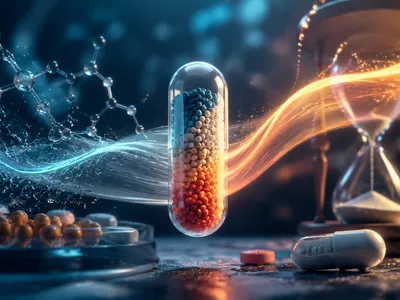
Pain Signal Origins
Tissue injury triggers the release of various biochemical mediators from damaged and immune cells, directly activating specialized peripheral nerve endings called nociceptors and converting harmful stimuli into electrochemical signals. Key mediators such as bradykinin, serotonin, and adenosine triphosphate (ATP) bind to specific receptor subtypes on nociceptive membranes, initiating intracellular signaling cascades that lower the neuron's activation threshold, a process known as sensitization. The enzyme cyclooxygenase converts arachidonic acid into unstable endoperoxides, which are then transformed into prostaglandins and thromboxane, amplifying pain signals at the injury site.
Prostaglandin E₂ (PGE₂) plays a major role in sensitizing nociceptors by activating EP receptors linked to G-protein pathways, increasing cyclic AMP levels, enhancing ion channel activity, and promoting more frequent neuronal firing. Inhibiting the production of these lipid mediators is the main mechanism behind many common analgesics, explaining why reducing prostaglandin synthesis effectively lowers both pain intensity and inflammation associated with tissue injury.
The following list summarizes the key molecular events from tissue damage to pain signal generation. Each step represents a potential target for pharmaceutical intervention.
- 🩹 Tissue damage releases arachidonic acid from membrane phospholipids via phospholipase A₂
- ⚡ Cyclooxygenase enzymes convert arachidonic acid to prostaglandin H₂ (PGH₂)
- 🧪 Tissue-specific synthases produce PGE₂, PGD₂, PGI₂, and thromboxane A₂
- 🔔 PGE₂ binds EP receptors on nociceptor terminals, lowering activation threshold
- 📡 Increased neuronal firing transmits pain signals to the spinal cord and brain
Each class of painkiller disrupts a distinct point in this signaling pathway. Nonsteroidal anti-inflammatory drugs (NSAIDs) target cyclooxygenase directly, while local anesthetics block voltage-gated sodium channels on the nerve fiber.
The specificity of these interventions determines their clinical efficacy and side effect profiles. Selective targeting of peripheral mediators offers pain relief with reduced central nervous system adverse effects.
COX Enzyme Role
The cyclooxygenase (COX) enzyme has two main isoforms with distinct roles. COX-1 is constitutively present in most tissues, supporting gastric mucosal integrity and platelet aggregation via thromboxane, while COX-2 is rapidly induced during inflammation, producing prostaglandins that drive pain, fever, and swelling at injury or infection sites.
Both isoforms share a similar catalytic pathway, converting arachidonic acid to PGG₂ through cyclooxygenase activity, then reducing it to PGH₂ via peroxidase. PGH₂ serves as the substrate for downstream synthases that generate active prostanoids, central to pain and inflammatory signaling. The discovery of constitutive and inducible isoforms shaped modern analgesic development.
Traditional NSAIDs nonselectively block both COX-1 and COX-2, providing pain relief but causing gastrointestinal side effects. Selective COX-2 inhibitors (coxibs) were designed to reduce gastric damage while preserving analgesia, though cardiovascular risks emerged from disrupting vascular prostacyclin. Acetaminophen uniquely offers strong analgesia despite weak COX inhibition, highlighting the importance of personalized medicine and genetic testing in understanding individual drug responses, and likely acts via a peroxidase-sensitive form of COX-2 in the central nervous system.
Understanding isoform-specific roles enables tailored analgesic choices: acute musculoskeletal pain may benefit from nonselective NSAIDs, whereas chronic inflammatory conditions often require COX-2 selective agents with cardiovascular considerations. Currently, researchers are investigating how artificial intelligence can accelerate drug discovery contributes to the development of COX-1 sparing strategies and prostaglandin receptor antagonists to separate therapeutic pain relief from homeostatic disruptions.
How NSAIDs Block Prostaglandin Synthesis
NSAIDs inhibit cyclooxygenase by competing with arachidonic acid for the enzyme's hydrophobic channel. This prevents formation of prostaglandin precursors like PGG₂.
Aspirin uniquely acetylates a serine residue in COX, causing irreversible inhibition lasting the platelet's lifespan. Other NSAIDs bind reversibly and require higher doses for sustained effect.
The carboxylate group of most NSAIDs interacts with arginine 120 at the COX active site entrance. Meanwhile the hydrophobic portion occupies the channel's upper segment, creating a dual interaction that stabilizes the enzyme-inhibitor complex and blocks substrate access.
Ibuprofen and naproxen exhibit similar binding kinetics, reaching peak inhibition within two hours but allowing enzyme recovery as drug concentrations decline. Flurbiprofen demonstrates higher potency through additional hydrogen bonding with tyrosine 355. The clinical consequence of reversible versus irreversible COX blockade determines dosing frequency and duration of action. Aspirin's permanent COX-1 inhibition in platelets underlies its unique cardioprotective effect, whereas reversible inhibitors require twice-daily dosing for sustained antiplatelet activity.
Ibuprofen and naproxen exhibit similar binding kinetics, reaching peak inhibition within two hours but allowing enzyme recovery as drug concentrations decline. Flurbiprofen demonstrates higher potency through additional hydrogen bonding with tyrosine 355. These variations illustrate how pharmacokinetics influence dosage and safety, especially since the clinical consequence of reversible versus irreversible COX blockade determines dosing frequency and duration of action.
The table below compares major NSAID classes according to their COX selectivity and clinical implications.
| NSAID Class | COX-1 Selectivity | COX-2 Selectivity | Key Clinical Note |
|---|---|---|---|
| Aspirin | High (irreversible) | Moderate | Cardioprotection at low doses |
| Ibuprofen | Moderate | Moderate | Short half‑life, OTC availability |
| Naproxen | High | Low | Longer duration, higher GI risk |
| Celecoxib | Very low | High (selective) | Reduced GI toxicity, cardiovascular caution |
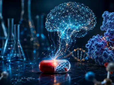
The Unique Brain Pathway of Acetaminophen
Acetaminophen is metabolically activated in the brain by fatty acid amide hydrolase (FAAH), generating AM404, an endogenous cannabinoid reuptake inhibitor. Unlike NSAIDs, it shows minimal peripheral anti-inflammatory effects, with analgesic action primarily in central nervous system regions such as the periaqueductal gray matter. AM404 activates TRPV1 channels and inhibits anandamide breakdown, increasing endocannabinoid levels and modulating descending pain pathways without affecting prostaglandins in peripheral tissues or the gastric mucosa.
Pharmacokinetic studies indicate that acetaminophen has an analgesic ceiling around 1000 mg, with no extra benefit at higher doses. Its sparing of COX‑1 ensures excellent gastrointestinal safety, while minimal COX‑2 inhibition limits anti-inflammatory effects. Emerging evidence also points to a secondary mechanism via the serotoninergic system, particularly 5‑HT₃ receptor antagonism, enhancing its efficacy in postoperative and chronic pain management.
Opioid Receptors and Pain Relief
Opioid receptors are G-protein-coupled proteins responsible for modulating pain and producing powerful analgesia without affecting consciousness. Activation of the μ-opioid receptor by morphine or naturally occurring endorphins inhibits adenylyl cyclase and blocks calcium channels, thereby reducing neurotransmitter release from presynaptic terminals in the dorsal horn and weakening pain signal transmission. Additionally, δ and κ receptors also play roles in analgesia, though they are associated with distinct side effects, as κ receptor activation may induce dysphoria while δ receptors influence the emotional perception of pain.
Chronic opioid exposure leads to receptor desensitization and downregulation, causing analgesic tolerance. The primary opioid receptor subtypes and their effects are listed below.
- μ (MOR): Supraspinal analgesia, euphoria, respiratory depression, and physical dependence
- δ (DOR): Spinal and supraspinal analgesia, reduced anxiety, minimal respiratory effect
- κ (KOR): Spinal analgesia, dysphoria, diuresis, and no respiratory depression