Vestiges of Our Ancestral Past
The human body functions as a living museum, preserving numerous anatomical features that have lost their original purpose through evolutionary change. These vestigial structures provide tangible evidence for common descent and adaptation over deep time.
While no longer critical for survival in modern environments, these remnants often reveal the specific selective pressures faced by our ancestors. The following list details some of the most prominent vestigial traits found in Homo sapiens, each telling a story of a different evolutionary pathway and environmental context that shaped our lineage.
- The palmaris longus muscle is absent in approximately 14% of the population, serving no significant function in modern grip strength.
- The coccyx, or tailbone, represents the remnant of a lost tail, providing minor support for pelvic muscles and ligaments.
- Goosebumps (piloerection) are a thermoregulatory reflex inherited from furry ancestors, now triggered primarily by strong emotions.
- The presence of a embryonic tail during fetal development is a potent recapitulation of our phylogenetic history.
The Pelvis and Bipedal Revolution
The hominin pelvis underwent profound morphological change to facilitate habitual bipedalism. This adaptation fundamentally distinguished our lineage from other primates.
A broader, bowl-shaped ilium reconfigured the attachment points for gluteal muscles, notably gluteus medius and minimus, which stabilize the torso during walking. This change narrowed the birth canal, creating an obstetric dilemma that drove secondary adaptations like altricial offspring.
Comparative analysis of hominin fossils reveals a clear trend in pelvic morphology correlating with increased bipedal efficiency. The table contrasts key features between an arboreal primate ancestor and modern humans, highlighting functional trade-offs. This skeletal compromise illustrates how a single selection pressure can reconfigure an entire anatomical module, with cascading effects on life history and social behavior.
| Pelvic Feature | Arboreal Primate (Ancestral Condition) | Modern Human (Derived Condition) |
|---|---|---|
| Ilium Shape | Tall and flat | Short, broad, and bowl-shaped |
| Sacrum Width | Narrow | Broad, with increased sacral curvature |
| Acetabulum Position | Laterally facing | More anteriorly (forward) facing |
| Primary Function | Arboreal locomotion, flexibility | Weight transfer, bipedal stability, birth canal formation |
The Compromised Human Spine
The human spine's characteristic S-shaped curvature is a direct but imperfect adaptation to bipedal locomotion, representing a significant departure from the single-arch spine of our quadrupedal ancestors.
While providing critical shock absorption and balance, this reconfigured column is prone to mechanical stress and pathological conditions rarely seen in other primates. The secondary lumbar curve, which brings the body's center of mass over the hips, places substantial compressive and shear forces on the intervertebral discs.
These biomechanical vulnerabilities manifest in widespread degenerative disorders. The prevalence of lower back pain and disc herniation is a modern eepidemic linked directly to our evolutionary history, a phenomenon described as evolutionary mismatch between our ancient anatomy and contemporary lifestyles. The following table compares spinal regions in humans and ancestral quadrupeds, highlighting the structural trade-offs that enabled upright walking at a cost. The cervical and lumbar lordoses are particularly susceptible to wear, revealing the spine as a structure still evolving under new pressures.
| Spinal Region | Primary Curvature (Quadruped) | Primary Curvature (Human) | Functional Consequence & Common Pathology |
|---|---|---|---|
| Cervical | Mild convexity (lordosis) | Pronounced lordosis | Supports head; susceptible to strain and degenerative joint disease. |
| Thoracic | Primary kyphosis (concave) | Kyphosis | Anchors rib cage; relatively stable but can develop hyperkyphosis. |
| Lumbar | Kyphosis | Pronounced lordosis | Bears most body weight; high incidence of disc herniation and spondylolysis. |
| Sacral | Kyphosis | Kyphosis (fused) | Transfers force to pelvis; sacroiliac joint dysfunction. |
Wisdom Teeth and Shrinking Jaws
The predictable impaction of third molars, or wisdom teeth, results from a pronounced evolutionary trend of masticatory apparatus reduction.
This reduction correlates with dietary shifts toward softer, processed foods and the controlled use of fire for cooking, which decreased selective pressures for robust jaws and large chewing surfaces. Consequently, the modern human jawbone is significantly smaller than that of early Homo or Pleistocene hunter-gatherers.
The discordance between genetically programmed tooth development and insufficient mandibular space creates a common clinical problem. This phenomenon is not merely a vestige but an active example of ongoing evolutionary change visible within historical timescales. The key factors driving this craniofacial reduction are summarized below, illustrating a complex interplay between behavior, technology, and anatomy.
- Dietary Shift: Transition from tough, fibrous foraged foods to softer, cooked diets reduced stress on jaw muscles and bones during growth.
- Technological Innovation: Tools for cutting, pounding, and later cooking externalized the mechanical processing of food.
- Genetic Changes: Mutations in regulatory genes like MYH16, associated with masticatory muscle size, may have relaxed constraints on jaw growth.
- Developmental Timing: The late eruption of third molars (ages 17–25) means they develop in a jaw that has already completed its growth, often leaving no room.
What is the Purpose of the Appendix?
Long dismissed as a classic vestigial organ, the appendix has undergone a significant reassessment in modern evolutionary medicine. No longer viewed as a pointless remnant, it is now understood as a complex secondary lymphoid organ that plays a role in the immune system.
This narrow, tube-like structure arising from the cecum is rich in gut-associated lymphoid tissue (GALT). It functions as a safe reservoir for beneficial gut bacteria, potentially enabling repopulation of the intestinal microbiome following a severe diarrheal illness. The evolutionary trade-off between this protective function and the risk of acute, life-threatening appendicitis presents a compelling example of antagonistic pleiotropy.
The changing perception of the appendix illustrates how a structure can retain functionality even as its primary evolutionary role shifts. The table below contrasts the historical view of the appendix with the contemporary understnding of its biological role, highlighting the nuanced reality of anatomical vestiges.
| Aspect | Historical View (Purely Vestigial) | Modern Understanding (Functional Vestige) |
|---|---|---|
| Primary Designation | Functionless remnant of a larger cecum for cellulose digestion. | Specialized component of the mucosal immune system. |
| Key Tissue | Simple columnar epithelium. | Concentrated gut-associated lymphoid tissue (GALT). |
| Postulated Function | None; an evolutionary "leftover." | Safe house for commensal bacteria; immune surveillance and training. |
| Evolutionary Context | Structure in the process of disappearance. | Structure with a repurposed, secondary adaptive function. |
Muscles We No Longer Command
The human muscular system contains several remnants that have lost their primary mechanical function, offering clear evidence of our ancestry. These muscles, often variable in presence and size between individuals, are controlled by the same nerves as their functional counterparts but produce no meaningful movement for modern humans.
The arrector pili muscles, which cause goosebumps, are a quintessential example. In furry mammals, they trap an insulating layer of air; in humans, they are vestigial reflexes triggered by cold or strong emotion. Similarly, the palmaris longus in the forearm is absent in about 14% of people and is clinically insignificant for grip strength.
Another notable vestige is the pyramidalis muscle, a small triangular muscle in the lower abdomen that tenses the linea alba. Its function is negligible, and it is absent in approximately 20% of the population. The persistence of these neuromuscular units, complete with nerve supply and embryonic origins, underscores their heritage from a functional past.
- Arrector Pili Muscles Universal
- Palmaris Longus ~86% of population
- Plantaris Muscle Variable
- Pyramidalis Muscle ~80% of population
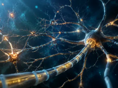
The variable expression of these muscles highlights the role of genetic drift in shaping modern anatomy once selective pressures are relaxed. Furthermore, some evolutionary theories, such as the axial twist hypothesis, propose that the very arrangement of our nervous system—with its characteristic contralateral organization—may itself be a deeply conserved anatomical legacy from early vertebrates.
This widespread contralateral control, where the left brain hemisphere governs the right side of the body and vice versa, is a fundamental but enigmatic feature of vertebrate neuroanatomy. While not vestigial in function, its near-universal presence across vertebrates suggests an ancient, conserved body plan feature whose evolutionary origin remains a subject of scientific investigation and may be linked to early asymmetric development.