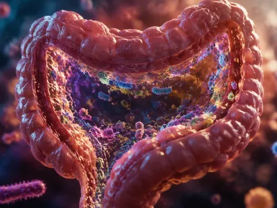
Microbial Immune Training
The neonatal gut is colonized by pioneer bacteria during birth, shaping immune tolerance by guiding regulatory T-cell development. Disruptions in this early microbial establishment can increase the risk of allergies and autoimmune diseases, highlighting that early-life microbiome is critical for setting immune balance.
Commensal bacteria such as Bifidobacterium and Bacteroides produce key molecules that activate immune receptors and stimulate anti-inflammatory cytokines, maintaining gut homeostasis. Without these microbial interactions, the immune system can become overreactive, leading to chronic inflammation.
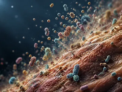
Gut Barrier Integrity
A single layer of intestinal epithelial cells forms a barrier between luminal microbes and host immune tissues, regulated by tight junction proteins. Mucus-degrading bacteria like Akkermansia muciniphila promote mucus production, strengthening this defense, while leaky gut allows bacteria to cross the barrier.
Barrier compromise permits microbial products such as lipopolysaccharide to enter circulation, causing systemic inflammation linked to metabolic endotoxemia, insulin resistance, and fatty liver disease. Butyrate-producing Firmicutes enhance tight junction assembly through AMP-activated protein kinase activation, reducing permeability and preventing excessive immune activation.
The following commensal genera play pivotal roles in preserving gut barrier function:
- Lactobacillus – reinforces tight junctions and produces antimicrobial peptides
- Faecalibacterium – generates anti-inflammatory butyrate
- Roseburia – strengthens mucus layer thickness
Commensal Metabolite Signals
Commensal bacteria generate short-chain fatty acids (SCFAs) like butyrate, propionate, and acetate. These molecules bind GPR41 and GPR43 receptors on colonic regulatory T cells.
Secondary bile acids deoxycholic acid and lithocholic acid modulate immune cell differentiation. They activate the nuclear receptor TGR5 on macrophages, reducing NLRP3 inflammasome activity.
Indole-3-aldehyde from Lactobacillus species activates the aryl hydrocarbon receptor in innate lymphoid cells and dendritic cells. This drives interleukin-22 secretion, which enhances epithelial integrity and antimicrobial defenses. Meanwhile, histamine produced by certain gut bacteria modulates histamine receptor H2 on dendritic cells, shifting cytokine profiles toward tolerogenic IL-10. Without these metabolite signals, the immune system fails to discriminate commensals from pathogens, leading to chronic mucosal inflammation and systemic autoimmunity, reflecting recent microbiology discoveries in human immunity.
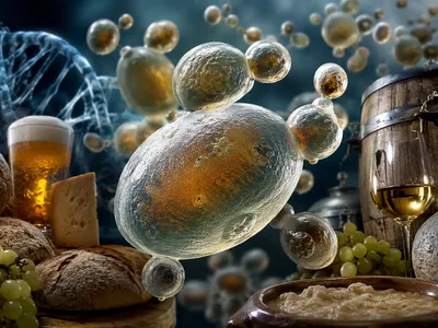
How Diet Reshapes Immune Function
Dietary fibers ferment into SCFAs, while dietary tryptophan fuels microbial indole derivatives. Both pathways calibrate intestinal immune responses and reinforce tolerance to food antigens.
High-fat, low-fiber Western diets reduce microbial diversity and deplete butyrate-producing Clostridia. This shift impairs regulatory T-cell induction and increases susceptibility to food allergies and colitis. Conversely, Mediterranean diets rich in polyphenols support anti-inflammatory commensals like Faecalibacterium prausnitzii, enhancing IL-10 production and gut barrier function.
Long-term dietary patterns alter the epigenome of immune cells via microbial metabolites. Butyrate functions as a histone deacetylase inhibitor, promoting Foxp3 expression in naive CD4+ T cells and suppressing pro-inflammatory interferon-gamma. Dietary vitamin A is converted by commensals into retinoic acid, which imprints gut-homing specificity on lymphocytes and enhances IgA production. A diet lacking fermentable substrates starves beneficial bacteria, causing overgrowth of pathobionts that trigger Th1-mediated tissue damage and systemic low-grade inflammation.
The table below summarizes how distinct nutritional components modulate commensal populations and subsequent immune functions. These interactions highlight diet as a central regulator of immune homeostasis.
| Dietary Component | Microbial Shift | Immune Consequence |
|---|---|---|
| High-fiber (resistant starch, inulin) | ↑ Bifidobacterium, Lactobacillus, SCFA producers | Treg expansion, increased IL-22, reduced Th2 responses |
| Saturated fats (lard, palm oil) | ↑ Bilophila wadsworthia, ↓ Roseburia | Th17 activation, gut permeability, endotoxemia |
| Polyphenols (flavonoids, tannins) | ↑ Akkermansia muciniphila, Faecalibacterium | Enhanced mucus barrier, reduced NF-κB signaling |
The Role of Bacteriophages in Immunity
Bacteriophages influence mammalian immunity through phage-encoded immunomodulatory proteins and activation of pattern recognition receptors. Gut phages accumulate at mucosal surfaces at high concentrations, engaging Toll-like receptors in dendritic cells and macrophages to trigger type I interferon responses without full inflammation. Phage transcytosis across epithelial barriers delivers viral particles to submucosal lymphoid follicles, priming adaptive immunity, and studies show that phages like Caulobacter crescentus phiCbK and Escherichia coli T4 reduce colitis severity in mice by modulating the NLRP3 inflammasome and enhancing regulatory T-cell populations.
In addition to direct immune modulation, phages support immune health by lysing pathogenic bacteria, lowering antigenic load, and limiting chronic immune activation. Phage-encoded endolysins break down bacterial peptidoglycan, releasing muramyl dipeptide that activates NOD2 and strengthens innate defenses. This combined action of immunomodulation and targeted bacterial killing underscores phages as key, yet often overlooked, regulators of immunity through their role in role of microbial communities in immunity.
- Myoviridae – induce type I interferons via TLR3 and TLR9 pathways
- Siphoviridae – promote regulatory T-cell expansion in gut-associated lymphoid tissue
- Podoviridae – reduce neutrophil infiltration and mucosal inflammation